What is a Bone Density Test?
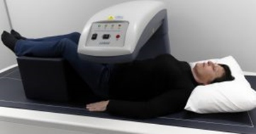
A Bone Density Test is also known as a Dual Energy X-ray Absorptiometry (DEXA or DXA). It is an enhanced form of X-ray technology that calculates bone mineral density which gives an indication of bone strength.
The purpose of a Bone Density Test?
It is used to diagnose osteoporosis, determine the risk of experiencing a minimal trauma fracture and monitoring of bone density. Osteoporosis involves a gradual loss of bone, as well as structural changes, causing the bones to become thinner, more fragile and more likely to break.
You may be referred for a bone density test if you are:
- over 70 years of age for screening of osteoporosis,
- you have previously been diagnosed with osteoporosis or osteopenia,
- on medications that may decrease your bone density, ie steroids – oral, topical, puffers.
- you have a disease that can decrease your bone density, ie liver, renal or thyroid disease and hormone disorders.
- Have suffered a minimal trauma fracture
How are the images acquired?
The DEXA scan is a non-invasive medical test that involves exposure of the body to a small dose of ionizing (X-ray) radiation. It sends two X-ray beams at different frequencies that target bone and soft tissue to produce an image so that the density of your bones can be determined.
Image courtesy of https://weightology.net
What happens during a Bone Density test?
On arrival, you will go through a questionnaire with a qualified technologist. This is to determine the appropriate images required and your risk factors for osteoporosis/ low bone density. It also helps us to determine any Medicare rebates that may be applicable.
Depending on what you are wearing, you may need to change into a gown. (Check below for preparation requirements)
Your height and weight measurements are taken just prior to the scan. This is required to be able to accurately perform your scan. (The weight limit of the scanner is 158kg.)
You then lie on the scanner and are positioned in the centre of the bed. Positioning is very important to get accurate results. The technologist will make sure throughout the procedure you are as comfortable as possible while at the same time we are able to obtain good, clear and accurate images.
Images of your hips and lumbar spine are taken routinely.
How long does the scan take?
The scan can vary between individuals and take from 10 to 20 minutes.
How do I prepare for the scan?
There is no preparation for the DXA scan. However, previous radiological procedures requiring a contrast injection (CT, MRI, Angiography and Nuclear Medicine) can interfere with the scan. When booking please let reception know if you have had any type of contrast in the previous 2 weeks so that an appropriate date can be booked.
It is also helpful but not essential to wear loose comfortable clothing without any metal (zippers, buttons, buckles or clips). Metallic objects interfere with the images. A gown is provided if clothing needs to be removed.
If you have had any spinal or joint surgery requiring any metallic implants (such joint replacements or screws) please let the technologist know and they will avoid scanning that area.
What are the risks of the scan?
The scan involves a very low dose of radiation. The radiation dose typically is less than a day’s natural background radiation.
- Bone Density (DXA) Spine and 1 hip 3-8μSv
- Body Composition (DXA) 0.5-4.5μSv
- Natural Background radiation per day 5-8μSv
- Lumbar Spine x-ray 700μSv
- Chest x-ray 20-150μSv
- Flight from Darwin to Perth 16μSv
As the dose is so low, this allows the technologist to be in the room with you throughout the whole procedure.
Are there any after affects for the scan?
There are no after affects from the procedure.
Are there any contraindications to the performing a Bone Density scan?
The scan IS NOT performed on pregnant women.
Any radiological procedures requiring a contrast injection 2 weeks prior to your scan can interfere with your results. When booking please let reception know so that an appropriate date can be booked.
Who does the scan?
A technologist who is qualified in Bone Mineral Density and holds a medical radiation license.
The images are then viewed by a qualified radiologist/physician and a written report is sent to your referring doctor.
The benefits of having the scan.
A bone density test is the only easily available test that can diagnose osteoporosis. This test helps to estimate the density of your bones and your risk/chance of breaking a bone. If osteoporosis or osteopenia is diagnosed, preventative measures can be started to help reduce the risk of future fractures and possible hospitalisation. Fractures to the hips and spine cause disability and may impede an independent lifestyle.
DXA is also effective in tracking over time the effects of treatment for osteoporosis and other conditions that cause bone loss.
To be able to track any significant changes in your bone density over a period of time, it is always best to have all follow up scans performed on the same scanner. Any changes in your calculated bone density will then be accurate and a true indication of any change.
Total Body Composition and VAT determination
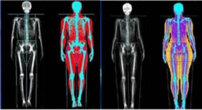
Body composition measures lean mass, fat mass, percentage fat and bone mineral content within the whole body as well various regions (arms, legs, trunk).
Scans can be useful in:
- Muscle weakness or poor physical functioning
- Limb asymmetry or wasting post-injury or surgery
- Endocrine imbalances
- Metabolic syndrome, assessment of visceral adipose fat (VAT)
- Weight loss regimens (help ensure you lose fat not muscle mass)
- After undergoing Bariatric surgery to assess fat and lean mass changes
- Assessing health risks associated with excess visceral fat
- Assessing for Sarcopenia in patients over 65years
- Monitoring weight disorders (obesity/metabolic disorders and anorexia)
- Sports performance (to ensure assessment/monitoring/setting safe targets for optimal health/performance in regard to correct fat/muscle ratio for your specific sport).
- Risk of Relative Energy Deficiency in Sport (RED- S)
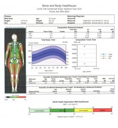
A body composition scan gives you:
- A colour mapped image of the whole-body showing fat mass, lean mass and bone
- percentage fat within the whole body and regional areas
- Lean muscle mass within the whole body and regional areas.
- Android/gynoid fat ratios
- Visceral fat
- Basal Metabolic Rate
- Relative Skeletal Muscle Index
- Bone mineral Density whole body (indication of osteoporosis risk)
- Changes can be tracked over time to ensure you are achieving your health, fitness or sporting goals.
- Fit Trace is available now to download and track your scan results via your smart phone https://www.fittrace.com
Visceral Adipose Tissue (VAT)
New software now means we can determine VAT (Visceral Adipose Tissue) within the android region of the whole-body scan. This is commonly known as “bad fat” as it surrounds internal organs such as the heart, liver and lungs. Visceral fat is associated with several health problems including Type 2-diabetes, heart disease, colon cancer and stroke. This can be used by referring clinicians for patient management either to treat these illnesses or lower your risk of getting them.
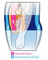
CoreScan results have been validated for adults between the ages of 18-90 and with a BMI (Body Mass Index) in the range of 18.5 – 40. Visceral Adipose Fat (VAT).
Sarcopenia (65 years and over)
Sarcopenia is a disease associated with the aging process. It is defined by loss of muscle strength and mass.
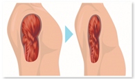
- Loss of muscle and strength can affect balance, gait and the ability to perform daily living tasks.
- Sarcopenia most commonly is seen in inactive people but can also affect individuals who remain physically active.
- When sarcopenia is coupled with other diseases associated with aging, its effects can be even more pronounced.
- Loss of muscle mass and strength is a significant risk factor for disability in the aging population.
- When patients suffer from both sarcopenia and osteoporosis, the risk of falling and subsequent fracture is higher.
Total body composition measures appendicular lean mass (ALM). When combined with values of muscle strength (grip strength, supplied by the referring doctor) and physical performance (gait speed, supplied by the referring doctor) a complete, integrated Sarcopenia assessment can be made.
Body Composition scans are useful when used in conjunction your Medical Practitioner, Accredited Practicing Dietitian, Sports and Exercise Physician, Physiotherapist, Accredited Exercise Physiologist (ESSA) or sports medicine doctor. It will help you and your health professional to monitor, track and assess your fat and lean mass changes, to improve your health, fitness and wellbeing.
This article was prepared by Bone and Body HealthScan.
